Dogma (noun) – A point of view or tenet put forth as authoritative without adequate grounds – Merriam Webster Dictionary
I did not set out to burn down the American Heart Association’s house, but I do go where the facts lead me and the facts do not match their dogma. If you have read “Cholesterol And Heart Disease” then you hopefully realize that cholesterol is not the cause of heart attacks or strokes and you are probably anxious to know the real cause. Ultimately the answer is simple, but we must first follow more facts to get there.
Risk Factors For Cardiovascular Disease (CVD)
The risk factors for CVD identified by the American Heart Association (AHA), the World Health Organization (WHO), the National Institutes Of Health (NIH), and the Centers For Disease Control and Prevention (CDC) are:
Elevated Cholesterol- Tobacco Use
- High Blood Pressure
- Type 2 Diabetes
- Obesity
- Physical Inactivity
I removed cholesterol from the list for obvious reasons. Additionally, the CDC and the NIH both list “diet” as a risk factor, by which they mean “cholesterol” so again I decline to include that. However, they also mean that one should eat better and I agree although I’m sure my list of approved foods is very different from theirs.
The CDC is the only organization to list “excess alcohol” as a risk factor. Technically that is true, but “excess” alcohol will kill you in any number of ways. Moderate alcohol consumption is, however, heart protective (this according to the AHA). I will leave alcohol off the list for now as it wasn’t included by the others.
Before I can begin tying these five generally agreed upon factors together we need to look at what happens before a stroke or heart attack.
A Closer Look At Arterial Plaque Formation
The Dogma: Plaque formation starts when you are about 10 and continues gradually over the years. Plaques are depositions of LDL.
The Problem: Gradual deposition of LDL into plaque fails to explain incidents of heart attacks in the young and otherwise healthy. It also fails to explain why plaque formation is not uniform along arteries instead of the discrete formation that takes place (i.e. plaques in different locations and of varying thicknesses). It also does not adequately explain how the lipids and clotting factors are able to move to the other side of the endothelium.
The Better Explanation: Plaques form in response to injury. The endothelium of the artery is damaged and bits are stripped away. The immune response includes clotting and a regrowth of the endothelium over the subsequent clot. The clot is eventually dissolved. This process of damage, clot, regrowth, and absorption is relatively common and if all goes well you’ll never notice. If, however, the damage is frequent enough to overwhelm the system you will start to have an enlarged area of unstable plaque that could rupture at any moment.
This explains why plaque formation is discrete, how clotting agents end up on the other side of the endothelium, and how plaque can grow rapidly during periods of acute injury to the endothelium. How LDL plays a role will be explained later.
Let’s fact check this hypothesis using research from the institutes of dogma.
Facts: From the American Heart Association journal Circulation:
The initial (type I) lesion contains enough
American Heart Association journal Circulation 1995atherogeniclipoprotein to elicit an increase in macrophages and formation of scattered macrophage foam cells. As in subsequent lesion types, the changes are more marked in locations of arteries with adaptive intimal thickening. (Adaptive thickenings, which are present at constant locations in everyone from birth, do not obstruct the lumen and represent adaptations to local mechanical forces). Type II lesions consist primarily of layers of macrophage foam cells and lipid-laden smooth muscle cells and include lesions grossly designated as fatty streaks. Type III is the intermediate stage between type II and type IV (atheroma, a lesion that is potentially symptom-producing). In addition to the lipid-laden cells of type II, type III lesions contain scattered collections of extracellular lipid droplets and particles that disrupt the coherence of some intimal smooth muscle cells. This extracellular lipid is the immediate precursor of the larger, confluent, and more disruptive core of extracellular lipid that characterizes type IV lesions. Beginning around the fourth decade of life, lesions that usually have a lipid core may also contain thick layers of fibrous connective tissue (type V lesion) and/or fissure, hematoma, and thrombus (type VI lesion). Some type V lesions are largely calcified (type Vb), and some consist mainly of fibrous connective tissue and little or no accumulated lipid or calcium (type Vc).”
The strikethrough is mine. Everything else is verbatim.
The same study reported 38% of advanced plaques had blood clots on the surface of the plaque and that an additional 29% of advanced plaques comprised layers of different ages containing elements of old blood clots. They conclude “the [clotting] that underlies thrombotic deposits in many cases may recur, and small thrombi reform many times. Repeated incorporation of small recurrent [clots] into a lesion over months or years contributes to gradual narrowing of the arterial lumen.,” (i.e. plaque forming in layers narrows the artery).
More facts about plaques:
“Initial fatty streaks evolve into fibrous plaques, some of which develop into forms that are vulnerable to rupture, causing thrombosis or stenosis. Erosion of the surfaces of some plaques and rupture of a plaque’s calcific nodule into the artery lumen also may trigger thrombosis. The process of plaque development is the same regardless of race/ethnicity, sex, or geographic location, apparently worldwide. However, the rate of development is faster in patients with risk factors such as hypertension, tobacco smoking, diabetes mellitus, obesity, and genetic predisposition.”
American Journal of Medicine – January 2009
Emphasis mine
Summary of the facts regarding arterial plaques:
- The AHA found plaques to contain lipids, white blood cells, and blood clots, often in layers.
- Fibrous plaques are more dangerous than calcified plaques because they are unstable and more prone to rupture.
- “The process of plaque development is the same regardless of
race/ethnicity, sex, or geographic location, apparently worldwide.” - The rate of development of plaques is not related to cholesterol or LDL levels. Believe me, if they had found a relationship with cholesterol it would have been shouted from the mountaintops.
Response To Injury and LDL
So far the response to injury hypothesis explains how the LDL would get to the other side of the endothelium, but not why it would end up there as part of the endothelial injury and recovery process.
We turn again to Dr. Malcolm Kendrick from his book The Great Cholesterol Con for an explanation that involves vitamin C, excess bleeding, and a little known apoprotein that has been used as a pretty good predictor of CVD.
We normally think of vitamin C as an antioxidant while forgetting its role in collagen formation and that is the key here. Collagen prevents blood vessels from becoming leaky. We know this because a severe lack of vitamin C leads to scurvy and the subsequent breakdown of collagen results in death by internal bleeding. Kendrick hypothesizes that during long periods when vitamin C was in short supply (like the ice age for example) a number of our ancestors with a genetic mutation for clotting were better able to survive. Specifically our ancestors who produced an apolipoprotein called Apo(a).
Apo(a) of course makes Lipoprotein(a) or Lp(a), which looks like all the other LDL floating around in the bloodstream, but importantly with apo(a) attached. I should note that the two terms are almost interchangeable so don’t get tripped up if we keep switching back and forth – just look for (a) – which by the way is called “little a” in casual conversation around the lab water cooler.G
Lp(a) has a strong affinity for areas of damaged endothelium where it rushes in to form a resistant clot. A normal clot contains an enzyme called plasminogen. When the time is right another enzyme (tPA) signals plasminogen to bust the clot from the inside out. Apo(a) mimics plasminogen and blocks the effectiveness of tPA, making the clot much more resistant to dissolving.
Apo(a) is interesting, but plaque contains LDL, right?
Unless you are specifically looking for apo(a) in plaque it is likely that all you will see is apoB and conclude that the plaque is 100% LDL, but studies that searched for apo(a) found it in high concentrations in atherosclerotic plaques.
We found that 83% of all apo[a] but only 32% of all apoB in lesions
Journal of Lipid Research 1991 – Cleveland Clinic
was in the tightly bound fraction. When normalized for corresponding plasma levels, apo[a] accumulation in plaques was
more than twice that of apoB. All fractions of tissue apo[a],
loosely bound, tightly bound, and total, correlated significantly
with plasma apo[a]. However, no significant correlations were
found between any of the tissue fractions and plasma apoB…. These
results suggest that Lp[a] accumulates preferentially to LDL in
plaques, and that plaque apo[a] is directly associated with
plasma apo[a] levels and is in a form that is less easily removable
than most of the apoB. This preferential accumulation of apo[a]
as a tightly bound fraction in lesions, could be responsible for
the independent association of Lp[a] with cardiovascular disease
in humans. -Pepin, J. M., J. A. O’Neil, and H. F. Hoff.
“Due to its strong genetic determination, Lp(a) levels are stable and are not significantly influenced by gender, age, or environmental factors…
Clinical Diabetes and Endocrinology 2016
The Emerging Risk Factors Collaboration have found that each 3.5-fold increase in Lp(a) resulted in a 13 % increase in CVD risk [21]. They also found that this association was continuous and become proportionally more important with higher Lp(a) levels. Moreover, they found that this association still persists even after correction for other lipid parameters“.
Emphasis mine.
Apo(a) is not only a better predictor of future CVD, the causal relationship is (unsurprisingly) independent of cholesterol level!
Cholesterol haters often point to Familial Hypercholesterolemia (FH) as evidence that cholesterol will kill you. It is true that individuals with FH are at elevated risk for CVD, but the evidence is mixed. Once again if cholesterol were the killer that the establishment believes it to be, few individuals with FH would live to a ripe old age (something that happens with alarming frequency if you’ve bet the farm on the cholesterol hypothesis). Interestingly, those with FH are much more likely to also have Hyperlipoprotein(a). This elevated apo(a) among FH is a much better explanation of increased CVD risk.
It turns out the response to injury hypothesis does explain how and why LDL ends up behind the endothelium and is so prevalent in plaque.
The Root Cause of Cardiovascular Disease
Now we have a viable explanation for how plaques form and why they contain cholesterol, but what causes the damage to the endothelium?
First let’s add a risk factor for heart disease that is often mentioned in conversation, but rarely makes it onto the sort of list put out by the AHA and others – stress. In some ways stress is harder to define or measure but it is real as are its effects. Kendrick differentiates between good stress and bad stress.
Good stress such as exercise (and other positive activities that raise the heart rate) and psychological stressors the create a healthy stress response (think performing well at school or work or your team winning) are not only not a concern, but are positive and necessary to good health.
Bad physical stress such as excessive, intense, forced exercise in adverse conditions, rheumatoid arthritis, cocaine use, smoking, eating under pressure, spinal cord injury, steroid use, diseases of the hormonal system and bad psychological stressors such as a bullying boss, suffering racism, money worries, your team losing and many many more are most definitely a concern. The list is long and detailed (my version is abbreviated) to illustrate how stress and endothelial damage is able to explain anomalies in epidemiological studies of cardiovascular disease that the cholesterol hypothesis simply cannot explain (i.e. the French paradox and other examples of high cholesterol populations with low levels of cardiovascular disease and studies showing sudden spikes in heart attacks in populations).
How does stress damage the endothelium?
Stress and the HPA-axis
HPA refers to the hypothalamus-pituitary gland-and adrenal glands. That anatomical combination works kind of like this – the hypothalamus sends a message to the pituitary gland which pushes hormones that act as messengers to the adrenal glands to produce steroid hormones. This HPA-axis is inseparable from the autonomic nervous system and its two parts: the parasympathetic and sympathetic nervous systems.
Fight or Flight
The sympathetic nervous system activates in times of stress and perceived danger. The signals that reach the adrenal gland primarily result in adrenaline and cortisol which speed up your heart rate, reduce saliva production, and redirect blood supply to the muscles. It also tells the liver to release its glycogen stores, which spikes glucose levels, and triggers blood-clotting factors. You are now better able to survive an encounter with the wild beast that has you cornered.
Relax and Eat
The parasympathetic nervous system does just the opposite. It slows your heart, stimulates insulin production and the release of bile, increases saliva, and sends blood to the intestines to aid digestion. There is no threat present so it is time to relax and enjoy a meal.
Fight-Relax/Run-Eat
Eating while stressed (an all too common occurrence) would be one example of how we often put these two systems into a deadly conflict. Instead of one or the other system sending signals, both would be putting out conflicting signals. The liver would be told both to release glycogen and to store it. Adipose tissue would be instructed to both absorb and release fats into the bloodstream. Blood would be called to and away from the intestines and muscles. Metabolic chaos would ensue. It’s not hard to imagine that this unbalancing of so many systems would lead to many health problems.
Is Stress Alone Damaging?
There are a many things going on with each system, but what if we cut to the chase and isolate a common stress hormone? Cushing’s disease is caused by a tumor in the pituitary that stimulates overproduction of a precursor hormone that in turn stimulates cortisol secretion from the adrenal glands. Those with Cushing’s disease are awash in cortisol.
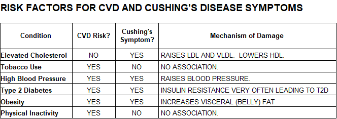
Cushing’s disease has a number of symptoms that look suspiciously like cardiovascular disease risk factors including one that I took off the list previous:

Cushing’s also raises at least four blood clotting factors. Remember the AHA’s list of plaque components? Clots and the remnants of clots were prominent.
Would it surprises you to learn that people with Cushing’s disease have accelerated plaque growth and a very large increase in their risk of heart disease?
You might also notice that Cushing’s changes cholesterol in ways that would alarm the AHA. This doesn’t bolster the AHA’s dogma so much as it indicates that, in some cases, changing cholesterol levels are likely a lagging indicator of atherosclerosis. To underscore the other more important factors of Cushing’s contribution to cardiovascular disease we need only look at the literature:
“In the clinical management of patients with CS [Cushing’s] the focus should be on identifying the global cardiovascular risk and the aim should be to control not only hypertension but also other correlated risk factors, such as obesity, glucose intolerance, insulin resistance,
Nueroendocrinology 2010.dyslipidemia, endothelial dysfunction and the prothrombotic state.” –
Strikethrough added.
Is cortisol really causing all these factors for heart attacks and stroke?
Cortisol also explains why steroids cause heart disease. Cortisol is the building block on which all steroids are built. Steroids are such a powerful immunosuppressant that they are used after organ transplants to prevent rejection and to treat conditions such as rheumatoid arthritis. Long term use of steroids increases heart disease risk. Even anabolic steroids, designed to increase muscle, can be damaging to the heart if overused.
“Compared with nonusers, anabolic-androgenic steroids (AAS) users … demonstrated higher coronary artery plaque volume than nonusers …. Lifetime AAS dose was strongly associated with coronary atherosclerotic burden …in rank of plaque volume for each 10-year increase in cumulative duration of AAS use.”
Circulation 2017
Admittedly, the above examples are extreme, so what happens when the HPA-axis suffers less extreme forms of unbalance?
- Depression – All of the Cushing’s related risk factors including deposition of visceral fat.
- Smoking – Increases cortisol and DHEA (another stress induced hormone) and increases blood clotting factors.
- Spinal Cord Injury – Severs most of the nerves for both the sympathetic and parasympathetic nervous systems. Symptoms are almost 100% matched to Cushing’s including greatly increased risk of CVD.
Relationship Between Factors Known To Damage The Endothelium and Cardiovascular Risk Factors
Let’s look again at the risk factors for cardiovascular disease as it relates to known factors for endothelial damage.

The other factors known to damage the endothelium include cortisol and high levels of adrenaline, cocaine, and acute mental stress.
From this information we can say with high confidence that stress increases stress hormones and that stress hormones (cortisol and adrenaline) damage the lining of the arteries and begin the response to injury process. The majority of other known risk factors for cardiovascular disease also damage the endothelium either directly or by increasing stress hormones.
The response to injury hypothesis explains why factors associated with damage to the endothelium can result in rapid or even sudden onset of heart attack or stroke. Some of you may remember the story of Len Bias, a 23-year-old basketball player who, after being drafted by the Boston Celtics, celebrated his success with cocaine and died of a heart attack. Given that the Celtics almost certainly performed exhaustive physical exams before offering a contract it is unlikely that Len Bias had any of the dogmatic risk factors for cardiovascular disease. I realize that this is a single case, but none the less illustrative of how massive endothelial damage can bring on a sudden and unexpected cardiovascular event.
Clotting And Its Outsized Role In Cardiovascular Disease.
Clotting factors are directly and consistently associated with increased risk of CVD. Clotting helps to form the plaque and a clot is the final step in the process that results in heart attack or stroke.
Factors Known To Increase Blood Clotting
- Fibrinogen
- Lp(a)
- VLDL
- And on and on
“Almost all coagulation proteins, including tissue factor, are found in atherosclerotic lesions in humans…. hypercoagulability, defined either by gene defects of coagulation proteins or elevated levels of circulating markers of activated coagulation, has been linked to atherosclerosis‐related ischemic arterial disease.”
Journal of Thrombosis and Haemostasis 2012
The List Of Drugs That Actually Reduce Your Risk of Heart Disease
- Aspirin
- Warfarin
- Alcohol
- tPA
- Statins
- Streptokinase
- Clopidogrel
- Ace-inhibitors
All of the above are anti-coagulants. Let me repeat, every drug listed above that has been shown to reduce you risk of heart disease is a “blood thinner.”
I have not hammered away at statins in this article, but please don’t interpret the above as an endorsement. Rather, to the degree statins work it is almost accidental. The lowering of cholesterol is not helping the situation (in fact it may very well increase your risk), while the anti-coagulant properties are just what you need. That said, there are cheaper and safer methods for thinning blood.
Other Drugs Prescribed For Heart Disease
- Beta-blockers: They block the effects of adrenaline (epinephrine).
- Calcium channel blockers: These treat high blood pressure.
- Potassium or magnesium: Both can reduce blood pressure.
Testing For Future Heart Disease
Cortisol Test – saliva is fine, but test must be repeated throughout the day. Typically we test cortisol at its peak in the morning, but if your HPA-axis is damaged you may read normal or even low for cortisol. The give-away is if your readings don’t change much during the day. Cortisol levels should decrease during the day, but a broken HPA-axis will often crank out the same levels of cortisol all day every day (like a shuffling zombie unable to walk or run). This means the HPA-axis has lost its ability to respond to circadian rhythms and changes in stressors.
Coronary Calcium Scan (CCS) or coronary artery calcium (CAC) scanning – a CT scan of the heart and arteries to measure the extent of calcified plaques. If plaques hang around long enough they eventually become calcified (the cause of arterial stiffening), which you may remember from earlier is not particularly dangerous because the plaque is now stable (not likely to burst and clot). So why is coronary artery calcification (CAC) strongly correlated with the rate of future cardiac events? Probably because calcification is the last stage of atherosclerosis and a CAC test showing extensive calcification strongly implies that there is an equivalent amount of fibrous plaque (unstable and prone to rupture and clotting) on its way to becoming calcified. The good news is that if your CAC test shows little or no calcification then you probably (at that moment) don’t have much plaque regardless of stage.
Factor V Leiden – a deficiency of factor V (five) results in excess clotting. This is genetic so you can find out if you are a “clotter” by taking a genome test such as 23andMe or (for about the same price) you can test for this one gene. For obvious reasons I would opt for the full test.
Preventing Cardiovascular Disease
What To Avoid
Avoiding these risk factors will reduce the frequency and severity of endothelial damage and diminish the aggressiveness of clotting in response to injury thereby reducing your risk of heart attack or stroke:

What To Do
Your proactive program looks like this:
- Start a ketogenic diet. It will help reduce stress, reduce high blood pressure, reverse obesity, and prevent or reverse type 2 diabetes.
- Meditate or find other forms of stress reduction and release
- Exercise. Cardiovascular or resistance or, better yet, both.
- Take a blood thinner if you have clotting issues. Consider at least a daily 81 mg aspirin as a preventative measure.
A Final Note or Two
For those of you wondering why a site dedicated to Alzheimer’s research has seemingly gone off the rails and become the site for heart attack advice consider this:
Of the nine risk factors for cardiovascular disease listed above all nine are also risk factors for Alzheimer’s, yes even vitamin C deficiency.
I can’t help but point out that reduced cholesterol levels are also a risk factor for rapid memory loss, confusion, and problems with forming new memories. This is important because statin makers have in the last few years tried to make the case that statins will prevent Alzheimer’s. I fear the opposite will be true.
A note on terminolgy – “chronic endothelial injury hypothesis (CEIH)” is not the same as response to injury. CEIH clings to the false notions that LDL magically appears beneath the intact endothelium and/or that LDL somehow damages the endothelium.
.

